
Introduction to Dental Exams & X-rays at Audubon Dental
Dental exams and X-rays at Audubon Dental in Thibodaux, LA are structured clinical evaluations used to assess the condition of teeth, gums, supporting bone structures, and surrounding oral tissues. These diagnostic procedures are foundational components of preventive and restorative dental care and are performed to document oral health status at specific points in time.
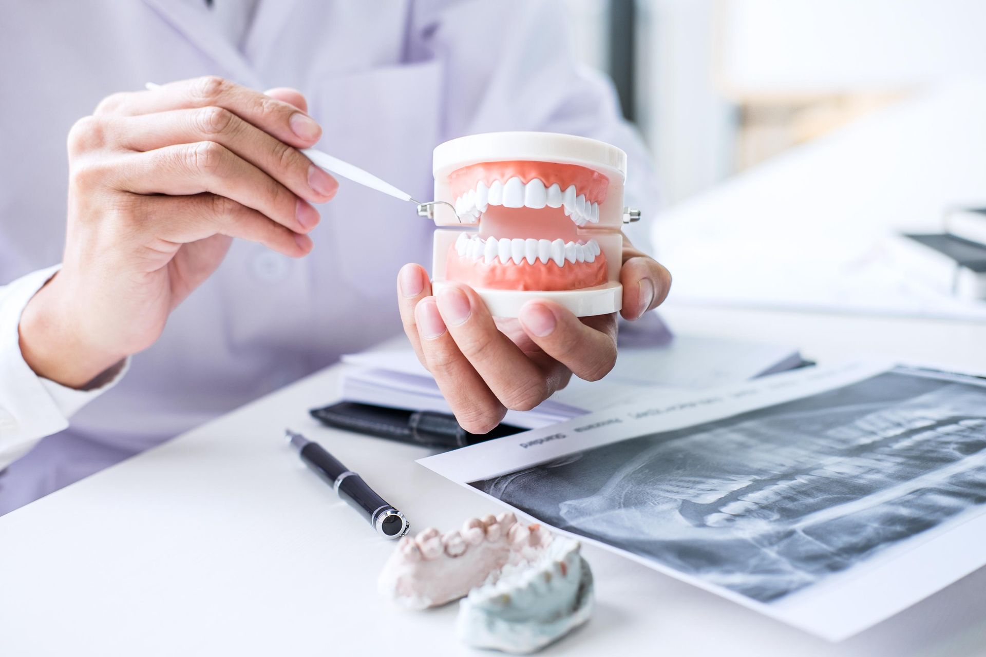
A dental exam typically includes both a visual and tactile inspection of the mouth, while X-rays provide internal imaging that cannot be observed during a standard clinical examination. Together, they create a comprehensive diagnostic profile that helps identify conditions affecting dental structures at early or advanced stages.
Exams & X-rays in Thibodaux, LA are scheduled based on patient history, oral health condition, and the need for ongoing monitoring of dental development or disease progression.
Clinical Purpose of Dental Examinations
A dental examination at Audubon Dental is designed to evaluate multiple aspects of oral health in a systematic and documented manner.
Visual Oral Assessment
During the visual assessment, the teeth are examined for structural integrity, wear patterns, discoloration, and surface irregularities. The gums are also inspected for inflammation, recession, and general tissue condition.
Soft Tissue Evaluation
Soft tissue structures such as the tongue, cheeks, palate, and floor of the mouth are reviewed for abnormalities. This includes checking for lesions, irregular textures, or changes in tissue appearance that may require further evaluation.
Occlusion and Bite Relationship Review
The alignment of the upper and lower teeth is assessed to determine how the bite functions during closure and movement. This includes identifying irregular contact points or uneven wear that may indicate functional imbalances.
These examination steps are recorded as part of the patient’s dental history and used as a reference for future evaluations in Thibodaux, LA.
Diagnostic Process During a Dental Exam
The diagnostic phase of an exam at Audubon Dental
combines clinical observation with data collected from imaging and patient history.
Review of Dental History
A review of prior dental records is conducted to understand past treatments, existing restorations, and previously identified conditions. This helps establish continuity in care and tracking of oral changes over time.
Periodontal Assessment
The condition of the gums and supporting structures is evaluated using clinical probing techniques. Measurements are taken to determine gum attachment levels and pocket depths.
Caries and Structural Integrity Evaluation
Each tooth is examined for signs of decay, cracks, or enamel erosion. Areas that present potential risk are documented for further investigation through radiographic imaging.
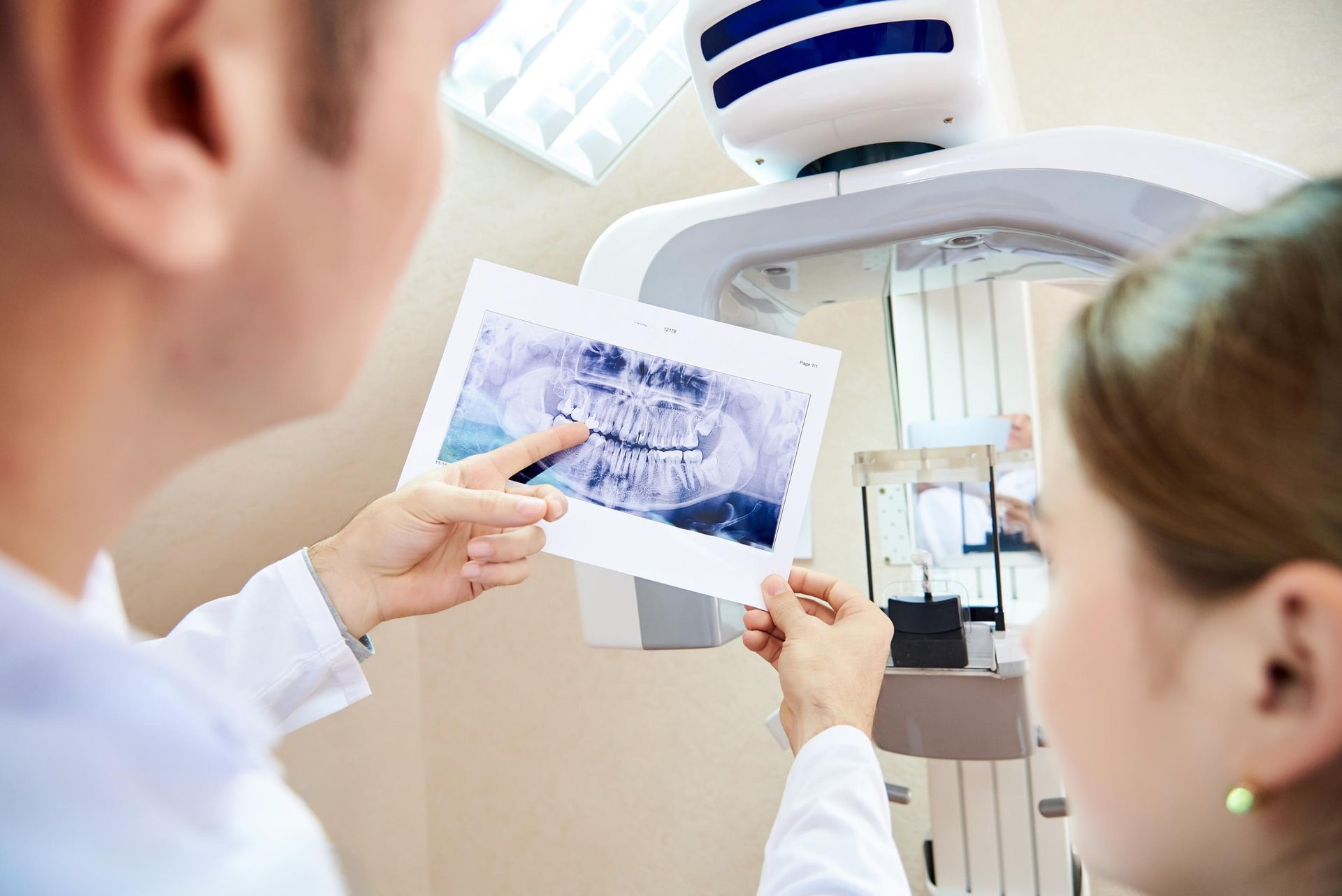
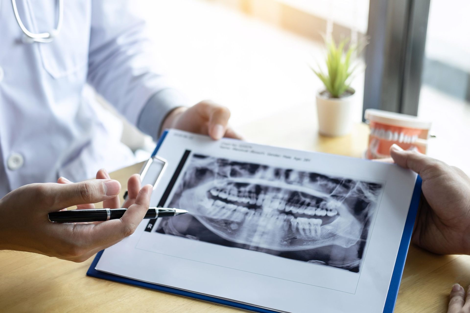
Dental X-rays and Imaging Procedures
Dental X-rays at Audubon Dental
in Thibodaux, LA are used to capture detailed internal images of the teeth and surrounding bone structures that are not visible during a clinical exam.
Bitewing X-rays
Bitewing images focus on the upper and lower back teeth and are commonly used to detect interproximal decay between teeth and evaluate bone levels supporting the teeth.
Periapical X-rays
Periapical radiographs capture the full length of individual teeth, including the root structures and surrounding bone. These images are used to assess root health, infection, or abnormalities at the tooth apex.
Panoramic X-rays
Panoramic imaging provides a broad view of the entire oral cavity, including all teeth, jaw structure, and surrounding anatomical features. This type of imaging is often used for comprehensive assessment and treatment planning.
Each type of X-ray serves a specific diagnostic purpose within the broader evaluation process performed in Thibodaux, LA.
Imaging Safety and Clinical Workflow
Dental imaging procedures at Audubon Dental
are conducted using controlled exposure protocols and standardized positioning techniques.
Radiation Exposure Control
X-ray equipment is calibrated to use minimal radiation necessary for producing clear diagnostic images. Protective measures are applied to limit exposure during imaging procedures.
Patient Positioning and Image Capture
Proper positioning is required to ensure accurate image quality. Sensors or film are placed in specific areas of the mouth depending on the type of X-ray being taken.
Image Review and Analysis
Once captured, radiographic images are reviewed for clarity and diagnostic value. The dentist evaluates the images alongside clinical findings from the oral examination.
This structured workflow ensures consistency in diagnostic imaging during exams & X-rays in Thibodaux, LA.
Documentation and Ongoing Oral Health Monitoring
Dental exams and X-rays at Audubon Dental contribute to long-term clinical documentation used to track changes in oral health over time.
Comparative Evaluation Over Time
Previous and current records are compared to identify changes in tooth structure, bone density, or gum condition. This comparison helps establish patterns of progression or stability.
Treatment Planning Integration
Findings from exams and imaging are used to inform future dental care decisions, including restorative, preventive, or periodontal procedures when needed.
Clinical Record Maintenance
All exam findings and X-ray results are stored as part of the patient’s permanent dental record in Thibodaux, LA. These records support continuity of care and ensure consistency in future evaluations.
Exams & X-rays at Audubon Dental function as a diagnostic framework that supports ongoing assessment of oral conditions through structured clinical observation and radiographic imaging.